
Anatomy Of The Foot Poster, Chiropractors, Osteopaths, Orthopedic Gift
Regions of the Foot. The foot is traditionally divided into three regions: the hindfoot, the midfoot, and the forefoot (Figure 2).Additionally, the lower leg often refers to the area between the knee and the ankle and this area is critical to the functioning of the foot.. The Hindfoot begins at the ankle joint and stops at the transverse tarsal joint (a combination of the talonavicular and.
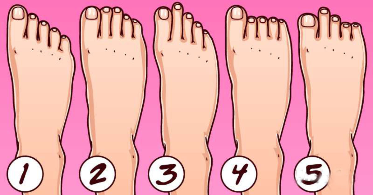
There Are 5 Types Of Feet Each Indicating A Particular Type Of Personality
Browse 19,300+ foot anatomy stock photos and images available, or search for foot anatomy vector to find more great stock photos and pictures. foot anatomy vector Sort by: Most popular Human foot anatomy cutaway representation, clipping path included. "Human foot anatomy cutaway representation, showing skin, veins and arterias, muscles, bones.

Labeled Diagram Of The Foot Teenage Lesbians
When to see a doctor Summary The foot is an intricate part of the body, consisting of 26 bones, 33 joints, 107 ligaments, and 19 muscles. Scientists group the bones of the foot into the.
Foot Pain Diagram exatin.info
Courtesy of Riley. Riley, a 21-year-old college student, sells feet pics online. She uses a "stage name" for anonymity. She didn't have time to get a second job, and she said feet pics are "pretty.
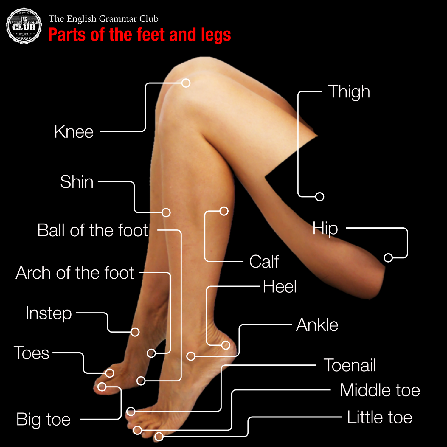
Parts of the feet and legs Grammar Tips
The ankle joint or tibiotalar joint is formed where the top of the talus (the uppermost bone in the foot) and the tibia (shin bone) and fibula meet. The ankle joint is both a synovial joint and a hinge joint. Hinge joints typically allow for only one direction of motion much like a door-hinge. Consequently, the ankle joint mainly only allows.

Vocabulary related to the foot for EFL students.. Learn english
486 human foot name stock photos, 3D objects, vectors, and illustrations are available royalty-free. See human foot name stock video clips Filters All images Photos Vectors Illustrations 3D Objects Sort by Popular Bones of the human foot with the name and description of all sites. Superior view. Human anatomy.
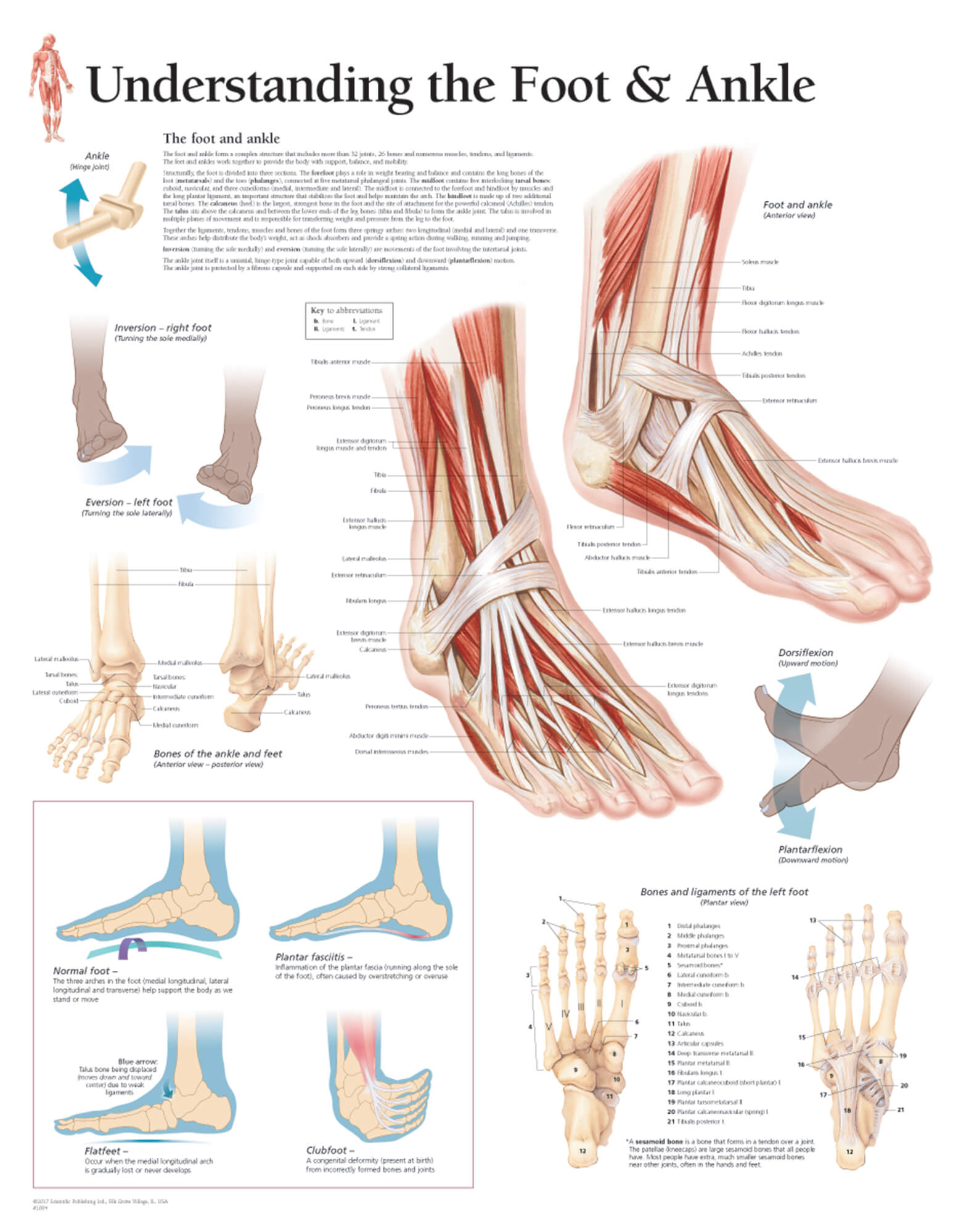
Understanding the Foot & Ankle Scientific Publishing
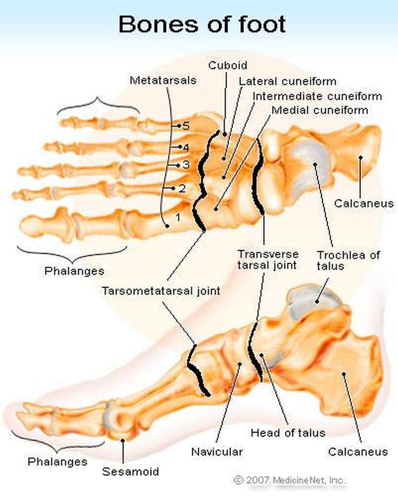
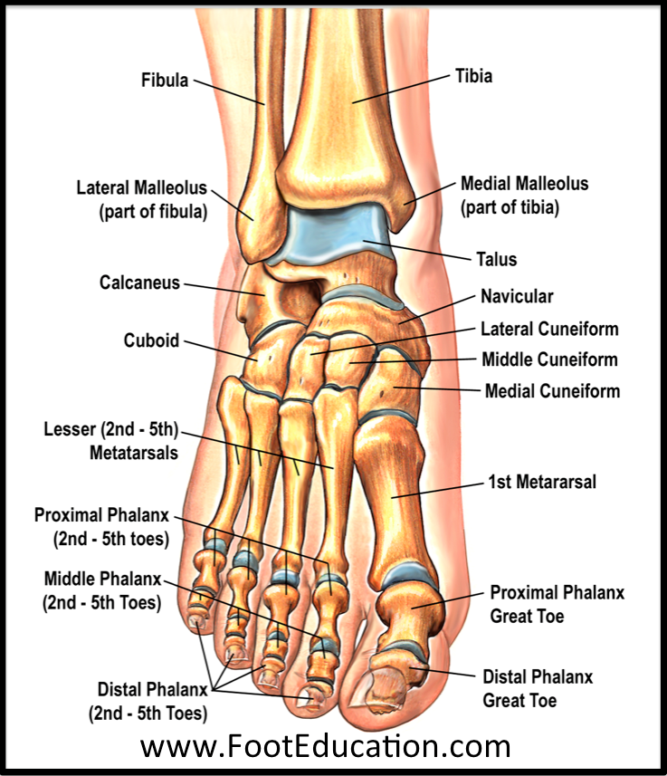
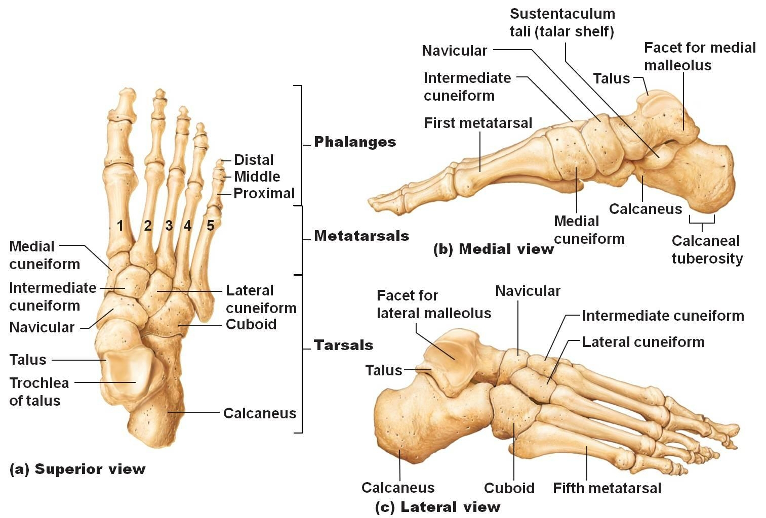
Bones of foot. The 26 bones of the foot consist of eight distinct types, including the tarsals, metatarsals, phalanges, cuneiforms, talus, navicular, and cuboid bones. The skeletal structure of.
Pictures Of Bones Of The Feet
Last updated 2 Nov 2018 The anatomy of the foot The foot contains a lot of moving parts - 26 bones, 33 joints and over 100 ligaments. The foot is divided into three sections - the forefoot, the midfoot and the hindfoot. The forefoot
Types Of Foot Problems Design Talk
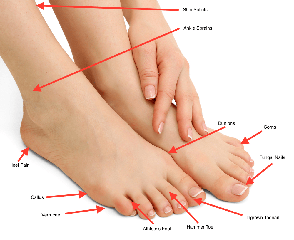
Stone bruise. Flat foot. Morton's neuroma. Sesamoiditis. Diabetic neuropathy. When to see a doctor. Foot conditions can vary in type, symptoms, and severity. These 17 issues are the most common. A.
Bones and Joints of the Foot and Ankle Overview FootEducation
Find Foot With Names Of Parts Of Foot stock images in HD and millions of other royalty-free stock photos, illustrations and vectors in the Shutterstock collection. Thousands of new, high-quality pictures added every day.
Lisfranc Injuries Core EM
F o o t B o n e s L a b e l e d D i a g r a m Names of the Bones in the Foot With Basic Anatomy Tarsal Bones The tarsals are a group of 7 irregular bones forming the hindfoot and the midfoot. These bones are arranged in two rows, proximal and distal. The bones in the proximal row form the hindfoot, while those in the distal row from the midfoot.

anatomy of the foot Ballet News Straight from the stage bringing
So, we recommend using Snapchat as part of your overall strategy to sell feet pictures, but always keep in mind its limitations and strengths. 10. FunwithFeet. FunwithFeet, a platform dedicated to.

the body foot A1 Learn english vocabulary, English vocabulary
Human Foot Anatomy royalty-free images. 50,888 human foot anatomy stock photos, 3D objects, vectors, and illustrations are available royalty-free.. Bones of the human foot with the name and description of all sites. Lateral view. Human anatomy. Vector illustration isolated on a white background. Human having pain in feet.3d illustration.

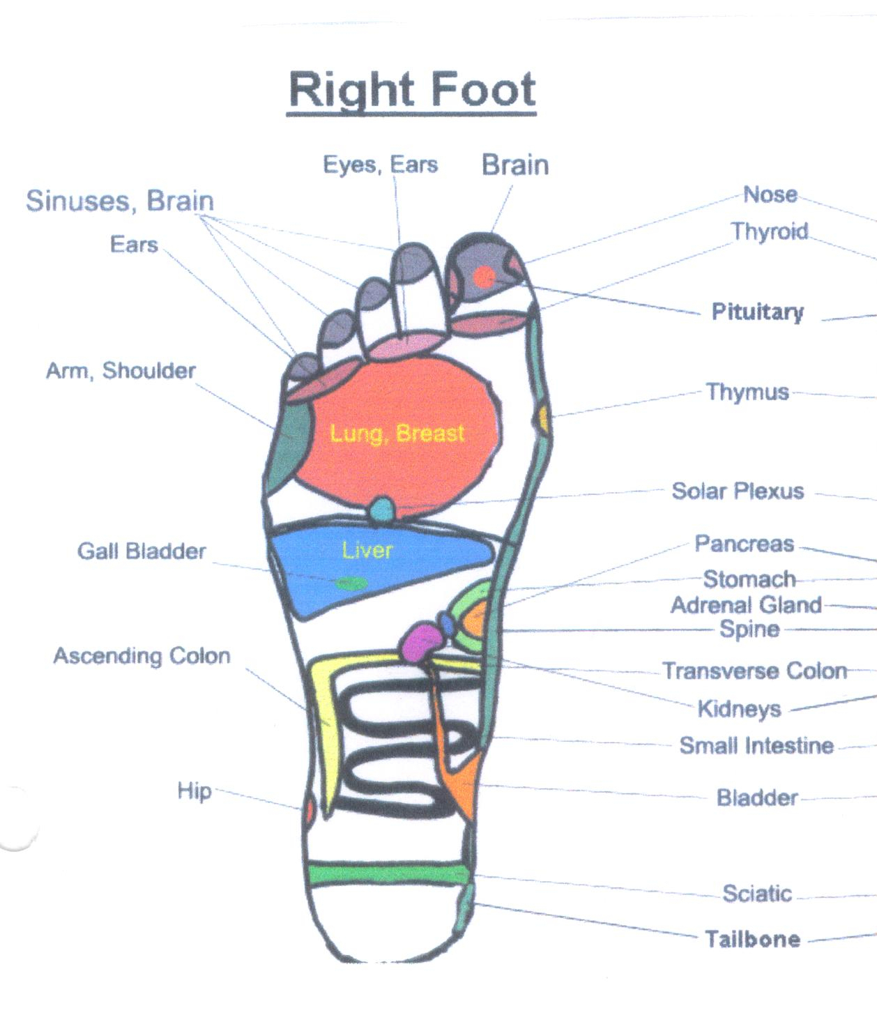
Anatomy Regions Of The Right Foot Wood Print lupon.gov.ph
Creative Editorial Human foot anatomy Stock Photos and Images (16,849) See human foot anatomy stock video clips Quick filters: Cut Outs | Vectors | Black & white Sort by Relevant RF PM67F9 - Human Foot Anatomy Illustration RF 2A13RHK - Vintage anatomy print of the human foot, showcasing the veins and arteries. RF D657GM - Foot anatomy, artwork
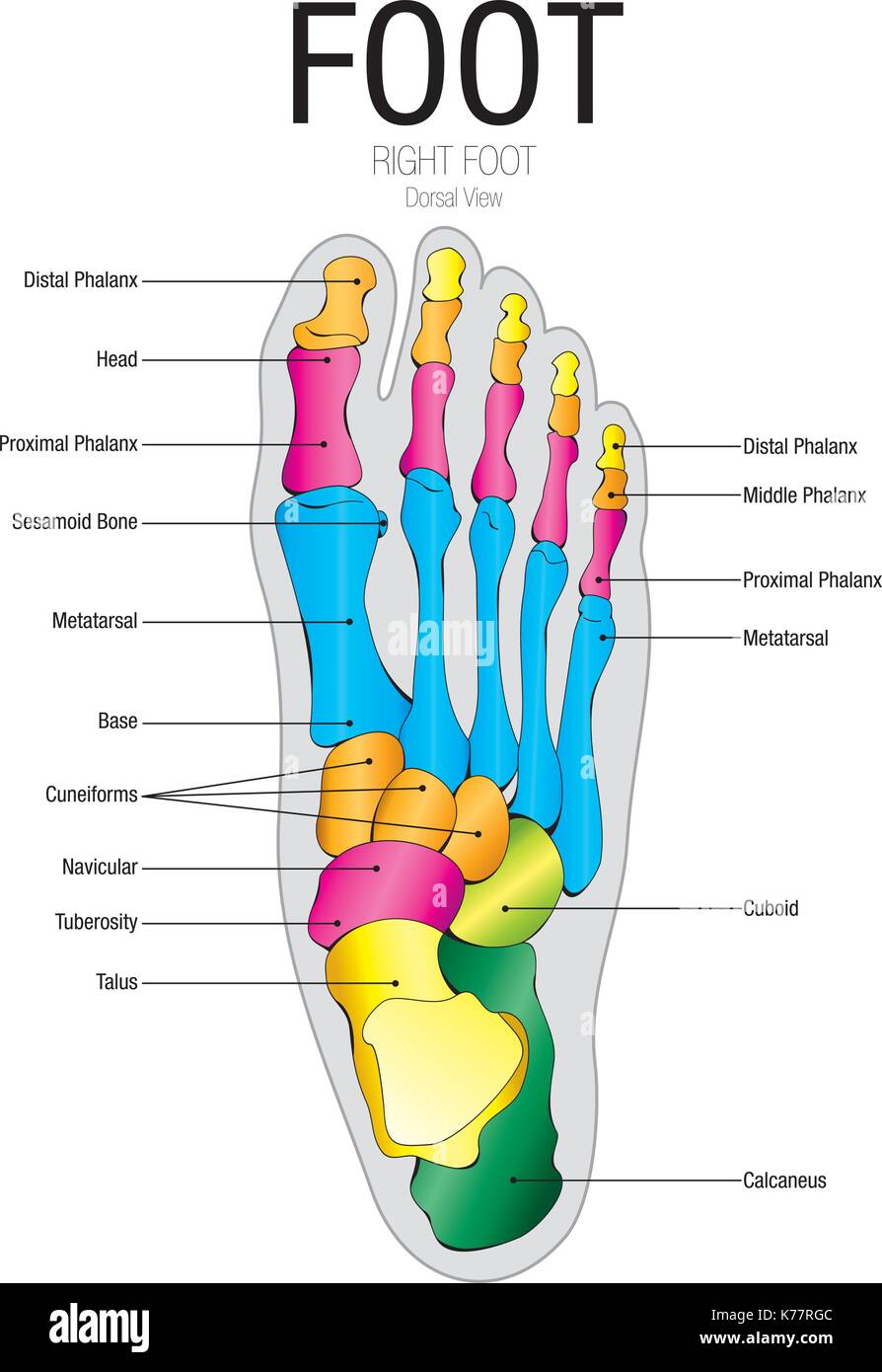
Chart of FOOT Dorsal view with parts name Vector image Stock Vector
Illustration Picture of Anatomical Structures - Foot Anatomy Picture of Foot The feet are located at the end of the legs and are used to stand and walk. Feet are very complex, comprised of 28 bones and 30 joints. The tendons, ligaments, and muscles in the feet number more than 100.
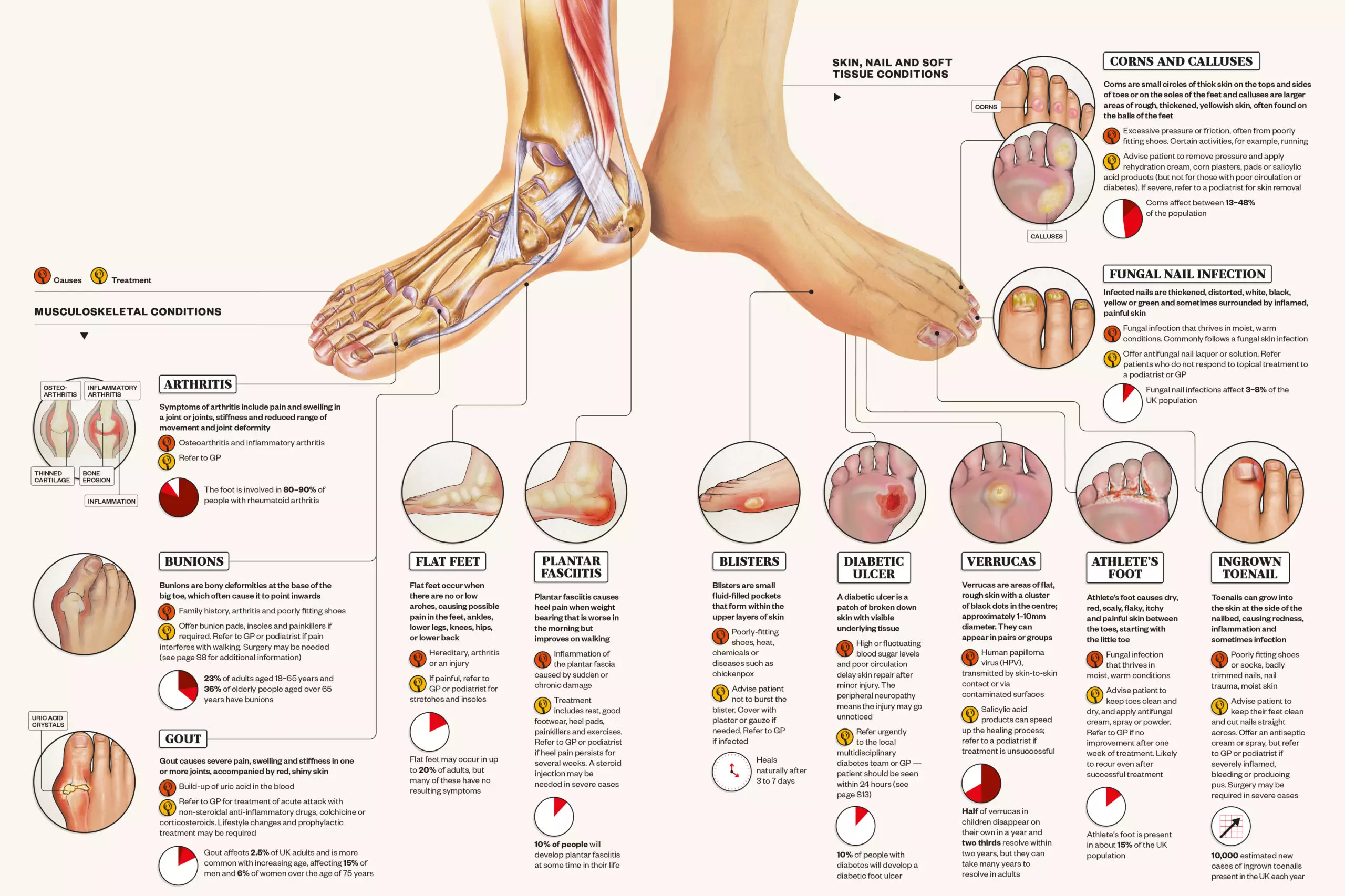
Identifying common foot conditions The Pharmaceutical Journal
Gout (a type of arthritis) Plantar fasciitis (heel pain) Stress fractures Diabetic foot ulcers Last medically reviewed on April 13, 2015 The foot is the lowermost point of the human leg.